Sound Solutions
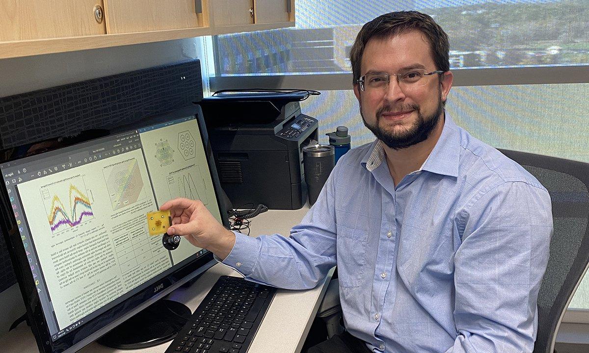
Dr. Wiacek’s laboratory, 医学声学全球健康成像和临床翻译(MAGIC)实验室, 继续加强SECS在生物医学成像和人工智能方面的专业知识.

Rehnuma Hasnat (M.S.)和本科生物工程学生Luc Taburet和莫莉Vue(不是图)博士一起工作. Wiacek博士参与了一个项目,该项目将人工智能与超声波和弹性成像相结合,以改善乳腺癌的诊断和筛查.
“Through my research, 我努力改进超声技术,为各种疾病的声学筛查方法创造一个新的范例,” says Alycen Wiacek, Ph.D., OU assistant professor, 谁在两个系之间工作——生物工程系和电气与计算机工程系. 当与用于信号检测的硬件和用于图像组合和显示的软件相结合时, 声学技术可以为新一代无痛手术铺平道路, ionizing radiation-free, and low-cost screening methods,” she adds.
Already as a Ph.D. student at Johns Hopkins University, Dr. Wiacek通过利用人工智能和其他先进的信号处理技术开发超声和光声成像系统,在提高诊断质量和患者护理方面取得了可观的进展. She implemented a novel deep learning architecture, 哪一个直接从原始超声数据中学习了基于物理的特征, 并创造了一种新的基于光声成像的手术指导系统,用于更安全的子宫切除手术.
In her second year at Oakland University, Dr. Wiacek’s laboratory, 医学声学全球健康成像和临床翻译(MAGIC)实验室, 继续加强SECS在生物医学成像和人工智能方面的专业知识.
One of her current projects, 由美国国家航空航天局通过密歇根太空资助联盟提供资助, 重点介绍了血栓形成的数字光学和声学模型的设计. More colloquially known as a blood clot, 该模型可用于确定定量图像特征的最佳组合,以随时间表征血凝块.
“In 2019, 在太空中发现一个凝块阻塞了宇航员的颈内静脉, 促使研究微重力对血栓形成的影响. In settings like the international space station, there are limited resources to make such a critical diagnosis. Ultrasound is the ideal imaging method for such detection, 但是标准亮度模式(b模式)超声不能随时间定量跟踪血凝块或描述它们的特征,” Dr. Wiacek explains.
Quantitative ultrasound, on the other hand, 能否利用组织的声散射特性来理解和表征组织微观结构的各个方面. Coupled with photoacoustic imaging, which leverages optical absorption properties of tissue, 它具有非侵入性地提供关于血凝块状态和组成的定量信息的潜力. Long term, 这些定量方法可以在离体和体内环境中实施, 能够在空间和地球上资源有限的情况下长期监测血栓形成.
Another research area that Dr. Wiacek目前正在研究将人工智能与超声波和弹性成像相结合,以改善乳腺癌的诊断和筛查.
Elastography, 它依赖于一个小的施加力之前和之后的组织运动的测量, is a promising method to complement standard B-mode ultrasound. 它可以评估组织内嵌块的刚度. However, b模式和弹性成像仍然受到噪声和伪影的影响,这些噪声和伪影是由波通过组织层传播引起的.
“通过在模拟超声和弹性成像图像中添加特定类型的噪声,我们可以模拟真实的临床成像场景. 然后我们通过给它噪声图像和干净图像来训练网络, 让它学习噪音是什么样子的,以及如何消除它. Consequently, when the network is given a new image, 它能够有效地去除噪声,提高整体图像质量.” says Dr. Wiacek.
“我们正在设计的神经网络利用了传统b超图像和弹性图的信息,以提高两种模式的图像质量,最终更好地可视化乳房肿块,” says Molly Vue, bioengineering undergraduate student, who works with Dr. Wiacek对该项目进行了研究,最近在2023 Sigma Xi国际卓越研究论坛上获得了工程学本科海报展示奖.
该研究的目标是创建模拟和模拟超声和弹性成像图像的数据集,并开发优化的网络架构以提高图像质量. At the conclusion of the study, Dr. Wiacek计划公开发布所有代码和数据集,以便将来进行基准评估. This study is funded by the OU University Research Committee.
“With proper engineering design, acoustic-based technologies are low cost, painless, and portable, making them the future of many disease screening solutions. Dr. Wiacek’s work is enabling this future. 通过使用多模态集成技术创建基于声学的筛选硬件, optimizing this hardware for seamless transition into the clinic, developing and optimizing algorithms for better image display, and building AI models, together, she is sculpting the future of equitable solutions,” states Shailesh Lal, Ph.D., Department of Bioengineering professor and chair.
Dr. Wiacek的研究与美国国家科学基金会(National Science Foundation)和国立卫生研究院(National Institute of Health)几个小组的任务密切相关. 有关她的研究和当前项目的更多信息,请访问实验室网站 www.secs.joyerianicaragua.com/~awiacek

 December 20, 2023
 By Arina Bokas

